
 English | EN
An AI model can achieve impressive results without ever having seen a real patient. In microwave breast imaging, this is not unusual. Before clinical measurements become available at scale, algorithms are often developed using numerical models, simulated electromagnetic data, or physical breast phantoms. These controlled environments make it possible to place a tumor at a known location, change its size, modify tissue composition, repeat measurements and generate labels with a precision that would be difficult to obtain in clinical practice.
For machine learning, this sounds almost ideal. But it also creates an important question. When an AI model learns from a phantom, what is it really learning? The answer depends not only on the algorithm, but also on how faithfully the phantom reproduces the complexity of the breast.
Why phantoms are so useful for artificial intelligence
Deep learning relies on examples. The more varied and representative those examples are, the better a model can potentially learn the relationship between microwave measurements and the underlying tissue configuration.
In emerging imaging technologies, however, large clinical datasets are difficult to obtain. Acquisition systems are still evolving, clinical studies are costly, and reliable annotations require medical expertise. Phantoms provide a practical intermediate step. They allow researchers to control parameters such as breast geometry, fibroglandular content, tumor presence, tumor position, tumor size, dielectric properties, antenna configuration, measurement frequency, etc.
For detection, this means that tumor and non-tumor examples can be generated under reproducible conditions. For localization, the exact target coordinates can be known. For characterization, the physical size and dielectric contrast of the inclusion can be controlled. This makes phantom data particularly attractive after the learning problem has been divided into detection, localization and characterization tasks.
A phantom can be physical or digital
The word phantom describes several different experimental realities.
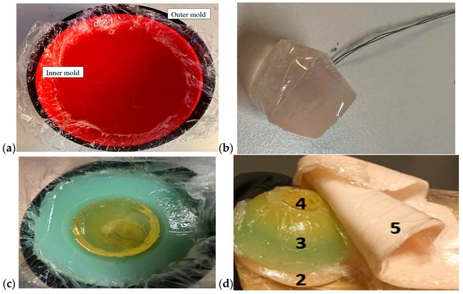
A physical phantom is a manufactured object containing materials designed to mimic the electromagnetic properties of biological tissues. Researchers can reproduce layers corresponding to skin, adipose tissue and fibroglandular tissue, then insert a tumor-like inclusion with controlled dielectric properties.
Recent work has demonstrated increasingly realistic three-dimensional tissue-mimicking platforms specifically designed for microwave sensing. Särestöniemi and colleagues, for example, developed breast phantoms containing separate skin, fat, glandular and tumor-mimicking regions and reported close agreement between target and measured dielectric properties.
A numerical phantom, by contrast, exists entirely in software. It represents the breast as a three-dimensional distribution of tissues to which dielectric properties can be assigned. Electromagnetic solvers can then simulate how microwave signals would propagate through that virtual anatomy.
Numerical phantoms offer another advantage: once the model exists, researchers can generate many controlled variations without physically manufacturing a new breast model for every experiment.
From simple geometry to anatomical realism
The earliest numerical experiments could rely on relatively simple shapes such as circles, ellipses or homogeneous layers. These simplified models remain useful for understanding algorithms, but they also make the learning problem easier than it will be in reality. A real breast contains heterogeneous fibroglandular structures, curved boundaries, skin, varying tissue proportions and potentially several lesions with different shapes and locations.
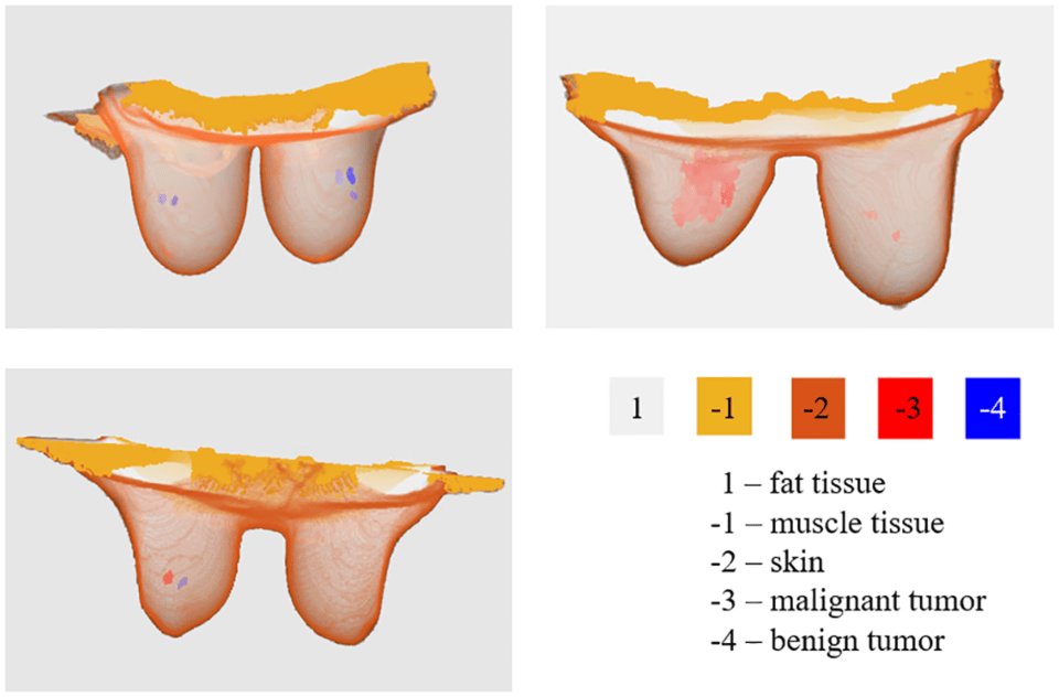
Recent datasets are therefore moving toward anatomically derived models. A 2024 open-access repository by Pelicano and colleagues provides three-dimensional breast models generated from clinical MRI examinations. The current repository described in the publication contains models derived from 55 patients, with normal breast tissues as well as benign and malignant tumors, including cases with multiple lesions. Dielectric property maps can be generated across the 3 to 10 GHz range.
This represents an important shift. Instead of asking only whether an algorithm works on an idealized breast, researchers can expose models to much more realistic anatomical variability.
Realism has more than one dimension
A phantom may look anatomically realistic and still produce unrealistic microwave data. This is because visual resemblance is only one part of the problem. For microwave imaging, at least four forms of realism matter.
- Anatomical realism: The geometry should represent realistic breast shapes and internal tissue distributions.
- Dielectric realism: Permittivity and conductivity should reproduce biologically plausible values across the frequency range used by the acquisition system.
- Electromagnetic realism: The measured or simulated signals should reproduce propagation, attenuation, reflections and scattering under conditions similar to the intended imaging system.
- Population realism: The dataset should contain enough diversity in breast composition, lesion position, lesion size and tissue properties to prevent the model from learning only a narrow family of experimental configurations.
The last point is particularly important for machine learning. A single very realistic phantom repeated hundreds of times does not automatically create a diverse dataset.
The danger of learning the experiment instead of the tumor
Consider a dataset where tumors always appear near the center of the phantom. A neural network could achieve excellent localization performance while implicitly learning that suspicious signals usually originate from the central region. Or imagine that tumor-containing phantoms were measured during one experimental session while healthy phantoms were measured during another. Small differences in calibration, temperature, antenna positioning or background noise could become correlated with the class label. The model might then learn the measurement session rather than the physical signature of the lesion.
This is one of the central risks of AI applied to controlled experiments. High accuracy does not necessarily mean that the intended phenomenon has been learned. Recent reviews of machine learning in microwave medical imaging repeatedly identify limited data availability, experimental variability and the gap between simulations, phantoms and clinical measurements as important barriers to robust translation.
Making phantom datasets harder on purpose
A useful training dataset should therefore introduce variability deliberately. Instead of keeping every parameter fixed except tumor presence, researchers can vary breast size and shape, tissue density, tumor position, tumor diameter, dielectric contrast, antenna positions, measurement noise, calibration conditions, frequency sampling, etc.
This changes the role of the phantom. It is no longer simply an object used to demonstrate that a tumor can be detected. It becomes a controlled generator of variability.
For artificial intelligence, this distinction is crucial. The objective is not to create the easiest possible learning problem. It is to create a learning problem that forces the model to identify features that remain meaningful when experimental conditions change.
Simulated data and experimental data should not be treated as identical
Numerical models offer scalability. Physical measurements offer experimental realism. Neither is sufficient on its own. Simulations can generate large amounts of perfectly labelled data, but the electromagnetic model cannot reproduce every imperfection of a real acquisition system. Physical phantoms naturally introduce measurement noise, antenna behaviour, coupling effects and calibration variability, but they are slower and more expensive to produce.
A promising strategy is therefore to combine different levels of data: numerical simulations → physical phantoms → clinical measurements. Each level introduces a new layer of complexity.
Deep learning studies are already exploring quantitative microwave imaging with large databases of anthropomorphic models, while experimental datasets provide an increasingly important test of whether the learned mapping survives outside simulation.
From controlled learning to credible learning
Phantoms are sometimes described as substitutes for patient data. A more useful way to think about them is as stepping stones toward patient data. They provide something that clinical datasets cannot easily offer at the beginning of a research programme: controlled conditions, repeatability and precisely known parameters.
But their scientific value depends on how they are used. A model that performs well across many anatomies, dielectric properties, lesion configurations and acquisition conditions provides stronger evidence than one evaluated on repeated measurements of a narrow experimental setup.
The next challenge is therefore not simply to generate more phantom data. It is to determine which aspects of the phantom should be considered ground truth, how reliable those labels really are, and how model performance should be validated when moving from controlled experiments toward clinical reality. That question deserves its own discussion.
Sources and further reading
- Särestöniemi, M. et al. Realistic 3D Phantoms for Validation of Microwave Sensing in Health Monitoring Applications. Sensors, 2024. https://doi.org/10.3390/s24061975
- Pelicano, A. C. et al. Repository of MRI-derived models of the breast with single and multiple benign and malignant tumors for microwave imaging research. PLOS ONE, 2024. https://doi.org/10.1371/journal.pone.0302974
- Shao, W. Machine Learning in Microwave Medical Imaging and Lesion Detection. Diagnostics, 2025. https://doi.org/10.3390/diagnostics15080986
- Silva, T. M. M. et al. Machine and deep learning applied to medical microwave imaging from reconstruction to classification. Journal of Physics Photonics, 2025. https://doi.org/10.1088/2516-1091/ae0bd3