 English | EN
Imagine a clinician trying to detect a tumor without relying on X-rays, MRI scans, or ultrasound images. Instead of looking at images, they analyze how electromagnetic waves travel through the breast. Tiny variations in those signals: reflections, phase shifts, and scattering patterns, can reveal the presence of abnormal tissue hidden beneath the surface.
This is the principle behind microwave breast imaging, a promising technology that aims to complement traditional breast cancer screening methods. Unlike mammography, microwave imaging relies on non-ionizing electromagnetic waves, making it potentially safer and more accessible for frequent monitoring.
But microwave systems do not start with images. They start with signals. And understanding how those signals become images is essential for research initiatives such as WaveCare, where artificial intelligence is being developed to detect, localize, and characterize tumors from microwave measurements.
Why microwaves interact differently with tumors
At the heart of microwave imaging lies a simple physical property: biological tissues have different dielectric characteristics. Breast tissues differ in their ability to store and conduct electromagnetic energy. Fatty tissue has relatively low permittivity, while fibroglandular tissue and tumors contain more water and exhibit higher dielectric constants. This contrast creates measurable differences in how microwave signals propagate through tissue.
When a microwave pulse travels through the breast:
- adipose tissue allows waves to propagate relatively smoothly,
- fibroglandular tissue causes moderate scattering,
- tumors often produce stronger reflections due to higher dielectric contrast.
These interactions leave subtle fingerprints in the measured signals. The challenge is to capture those fingerprints and interpret them.
Capturing the signals with S-Parameters
To measure these electromagnetic interactions, microwave imaging systems rely on scattering parameters, commonly called S-parameters.
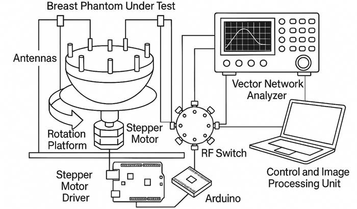
S-parameters describe how electromagnetic waves behave when they encounter a material or structure. In microwave engineering, they are the standard way to characterize signal reflection and transmission. They are typically measured using a Vector Network Analyzer (VNA) connected to antennas positioned around the breast.
Two parameters are particularly important in microwave imaging: – reflection coefficient (signal returning to the transmitting antenna) and
– transmission coefficient (signal traveling between antennas).
In practice, microwave imaging systems transmit signals across a range of frequencies while measuring both reflected and transmitted waves. The resulting dataset contains hundreds or thousands of signal measurements describing how the electromagnetic field interacted with the breast. But these measurements are still just numbers. Turning them into images requires solving a much harder problem.
From electromagnetic Signals to Spatial Images
Transforming microwave signals into images involves solving an inverse scattering problem. Instead of directly observing tissue structure, we observe the signals that emerge from it. The goal is to infer the internal dielectric distribution that could have produced those signals. This requires modelling how electromagnetic waves propagate through complex biological media.
A typical microwave imaging pipeline includes three main stages.
- Signal acquisition: Antennas transmit microwave signals while recording reflections and transmissions across multiple frequencies and positions. Modern systems often collect hundreds of measurements during a single scan.
- Electromagnetic modelling: Using Maxwell’s equations, reconstruction algorithms simulate how electromagnetic waves propagate through tissue with different dielectric properties. These models estimate how variations inside the breast would affect the measured signals.
- Image reconstruction: Finally, computational algorithms reconstruct spatial maps of dielectric properties. Common reconstruction techniques include delay-and-sum beamforming, confocal microwave imaging, inverse scattering tomography. If multiple scattered signals converge on the same spatial location during reconstruction, that region may correspond to a dielectric anomaly, potentially a tumor.
Why microwave image reconstruction is hard
Although the basic concept is intuitive, microwave imaging reconstruction is a notoriously difficult computational problem. Several factors contribute to this complexity.
- Multiple scattering: Microwave signals scatter repeatedly as they propagate through heterogeneous tissue, producing nonlinear interactions.
- Unknown tissue properties: Each patient has unique dielectric characteristics, which are rarely known beforehand.
- Limited measurement geometry: Antennas can only measure signals from specific locations around the breast, leaving gaps in the data.
Together, these challenges make microwave reconstruction an ill-posed inverse problem, meaning that multiple internal tissue configurations may produce similar measured signals.
The growing role of Artificial Intelligence
This is where artificial intelligence is beginning to transform microwave imaging research. Rather than solving the full physics-based reconstruction problem, machine learning models can learn patterns directly from microwave measurements or reconstructed images.
Deep learning models are increasingly explored to detect tumors (tumor vs. no tumor classification), localize tumors in 2D or 3D space, estimate tumor size and characteristics, tomography reconstruction. Instead of replacing physics-based methods, AI can complement them by creating hybrid pipelines where electromagnetic modeling and data-driven learning reinforce each other.
Signals Today, Clinical Insights Tomorrow
Microwave imaging challenges our traditional understanding of medical imaging. In most imaging technologies, clinicians start with images and interpret what they see. In microwave imaging, we start with signals. Those signals (tiny variations in electromagnetic waves) contain hidden information about tissue structure. Extracting that information requires a multidisciplinary effort combining electromagnetics, signal processing, inverse problem theory, artificial intelligence.
As these disciplines converge, microwave imaging may evolve from an experimental technology into a practical tool for early breast cancer detection and monitoring. And it all begins with something surprisingly simple: how a signal bounces back.
Sources and further reading
- Fear, E. et al., Microwave imaging for breast cancer detection? IEEE Potentials, 2003. https://doi.org/10.1109/MP.2003.1180933
- Khalid, N. et al., Emerging paradigms in microwave imaging technology for biomedical applications: unleashing the power of artificial intelligence. Nature, 2024. https://doi.org/10.1038/s44303-024-00012-8