
 English | EN
For microwave imaging, a machine learning model can perform extremely well in the laboratory. The antenna geometry is known. The breast models are controlled. The frequency range is fixed. Measurements are carefully calibrated. Training and test data come from the same experimental environment.
Then one detail changes. A different phantom is used. The acquisition frequency moves outside the training range. The antenna configuration changes. Measurements are collected with another system. Eventually, the data no longer come from a numerical model or a controlled phantom, but from a real patient. The patterns seen by the algorithm are no longer exactly the same. Nothing is necessarily wrong with the model. The data have simply moved into a different domain.
This phenomenon, commonly referred to as domain shift, is one of the major challenges facing medical artificial intelligence. In microwave imaging, it is particularly important because measured signals depend not only on tissue properties, but also on acquisition geometry, antennas, frequencies, calibration, coupling conditions and the many imperfections of a real measurement system.
The challenge is therefore no longer only to teach an algorithm how to recognize a tumor. It is to make sure that what the algorithm has learned still makes sense when the experimental environment changes.
When the data distribution changes
Machine learning models are usually trained under an implicit assumption that training and future test data follow similar statistical distributions. In a controlled experiment, this assumption may be approximately true. The same imaging system can be used repeatedly. The number of antennas remains fixed. The measurement protocol does not change. Similar phantom families may be used for both training and testing.
But microwave signals are highly dependent on the conditions under which they are acquired. A change in any of the following parameters can alter the measurements: breast geometry, tissue composition, tumor position and size, antenna arrangement, operating frequency, coupling medium, calibration procedure, measurement noise, instrumentation.
The biological phenomenon may remain the same while the measured S-parameters change. This distinction is essential. A model does not directly observe a tumor. It observes the electromagnetic consequences of a tumor through an acquisition system. If the acquisition environment changes sufficiently, the relationship learned during training may no longer hold in exactly the same way.
Recent reviews of medical AI describe this type of distributional difference between development and deployment data as one of the central barriers to robust model generalization.
The gap between simulation and experiment
Simulation is an attractive starting point for microwave imaging AI. Numerical models make it possible to generate large datasets while controlling anatomy, dielectric properties, tumor location, frequency and acquisition geometry. They also provide exact labels.
For deep learning, this is extremely valuable because experimental microwave datasets remain relatively small. A recent review of machine learning in microwave medical imaging notes that many published models still rely on only a few thousand simulated examples or tens to hundreds of experimental examples, which creates a significant risk of overfitting.
But numerical measurements are still models of reality. In simulation, antennas can behave exactly as defined. Cable losses can be ignored or accurately prescribed. Geometry can be perfectly known. Noise can be introduced according to a chosen statistical model.
Physical measurements are different. They contain antenna imperfections, cable responses, positioning errors, environmental interference, manufacturing tolerances and calibration inaccuracies. A simulated microwave signal is therefore an approximation of an experiment, not a duplicate of it.
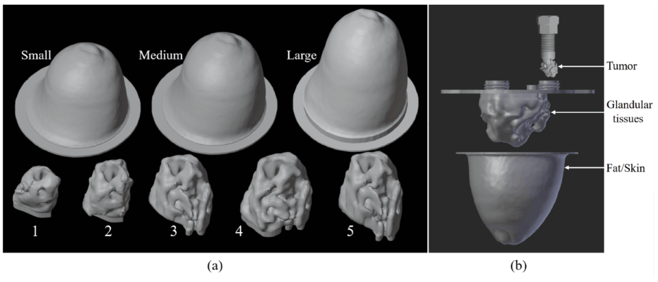
This kind of configurable dataset is valuable precisely because researchers can deliberately introduce variation into the training examples.
However, the same publication also highlights an important issue. Even when a dataset contains many combinations, training and validation samples may remain correlated if they are derived from common underlying phantom structures. The authors therefore use a separate phantom for testing generalization.
This illustrates a broader principle. Dataset size and dataset diversity are not the same thing. Thousands of examples generated from a narrow family of configurations may still describe a relatively small domain.
From one phantom to another
Physical phantoms reduce part of the simulation gap because they introduce real electromagnetic measurements. Signals now contain actual antenna responses, coupling effects and measurement noise. But another domain shift can still appear.
A model trained on one phantom family may encounter another phantom with a different external shape, another distribution of fibroglandular tissue, different dielectric properties, another skin thickness, different tumor positions, different manufacturing tolerances. This matters because deep learning models are extremely effective at finding statistical regularities.
The difficulty is that those regularities are not always the ones researchers intended them to learn. For example, if tumors are systematically placed within a narrow spatial region during training, a localization model may partly learn where tumors are usually placed rather than how their electromagnetic signatures encode location.
If tumor and non-tumor measurements are produced during different acquisition sessions, the network may exploit subtle differences in calibration or background noise. The algorithm can therefore learn the experiment instead of learning the phenomenon.
This is sometimes described more broadly as shortcut learning, where a model uses an easier correlated signal instead of the intended causal information. In a laboratory, such shortcuts can produce excellent test scores. Outside that laboratory, they may disappear.
When the acquisition system becomes part of the dataset
Microwave imaging introduces another important difficulty. The hardware itself strongly influences the data. A dataset collected with a monostatic scanning system is not statistically equivalent to one produced by a multistatic antenna array. Changing the number of antennas changes the dimensionality and spatial diversity of the measurements. Changing the frequency range modifies how waves interact with tissue. Changing the coupling medium affects wave propagation. Even differences in antenna design can alter amplitude and phase responses.
In other words: the acquisition system does not simply observe the dataset. It partly defines it. This has important consequences for AI modelling. A neural network trained on one particular representation of S-parameters may implicitly encode assumptions about the number of transmitting and receiving antennas, antenna ordering, frequency sampling, signal normalization, spatial geometry, system specific artefacts, etc. These assumptions may be invisible while development and testing use the same setup. They become visible when the model is transferred elsewhere.
A useful experimental example comes from previous work in three-dimensional microwave breast imaging, where a neural network trained on synthetic reconstructions was later tested with experimental data. The network remained useful when experimental frequencies were close to those represented during training, but failed to detect the target when measurements were obtained far outside the training frequency range. This offers a concrete illustration of how a seemingly small change in the acquisition domain can affect generalization.
The broader issue continues to appear in recent microwave AI literature. Current reviews identify data diversity, cross setup robustness and the transition between simulated and experimental measurements among the challenges that must be addressed before machine learning models can become reliable components of medical microwave systems.
The much larger step toward patient measurements
Moving from phantoms to patients introduces another level of variability. Even highly realistic phantoms remain deliberately controlled. Human anatomy is not. Breasts vary in size and geometry, adipose content, fibroglandular density, skin properties, internal tissue organisation, lesion morphology, physiological state, etc.
Patient positioning also introduces variability. Two acquisitions from the same person may not reproduce exactly the same geometry. These differences matter because microwave scattering is sensitive to both dielectric properties and spatial organisation. A neural network trained primarily on idealized or standardized geometries may therefore encounter signal patterns that were never adequately represented during training.
The problem is not exclusive to microwave imaging. Across established medical imaging modalities, studies consistently show that changes in scanners, protocols, institutions and patient populations can reduce the performance of deep learning models. A 2026 experimental study across radiological modalities found that models tested on images produced by different scanners generally performed worse than when evaluated within the same scanner domain.
For microwave imaging, where acquisition architectures are currently less standardized than technologies such as CT, the need to understand these shifts is particularly relevant.
Testing generalization rather than memorization
A random train and test split is useful, but it does not necessarily answer the most important question. Suppose a dataset contains repeated measurements from the same set of breast phantoms. Randomly assigning individual measurements to training and testing can place very similar examples on both sides. The model then appears to generalize, but the test environment remains closely related to its training environment.
Stronger evaluation protocols deliberately increase separation. A model can be tested on unseen tumor positions, unseen tumor sizes, unseen breast geometries, independent phantom instances, independent measurement sessions, additional noise conditions, different frequency samples, different acquisition configurations. Eventually, the test may involve another imaging system or an independent clinical site.
Each step answers a progressively harder question. The first asks whether the model has learned the training data. The last asks whether it has learned something that survives changes in the environment.
Modern medical imaging research increasingly distinguishes these questions through domain generalization, where models are expected to work on previously unseen domains, and domain adaptation, where information from a new target domain is used to adapt the trained model.
Making variation part of the training strategy
One response to domain shift is to make the training environment less predictable.
- Data augmentation: Artificial variation can be introduced into training data. For microwave signals, this may involve modifying noise levels, signal amplitude, phase, frequency sampling or geometrical parameters. The objective is to discourage the network from relying on fragile patterns that only exist under one acquisition condition.
- Domain randomization: Domain randomization pushes this principle further. Instead of trying to reproduce one experimental system perfectly, many parameters are intentionally varied during simulation. A training database could vary antenna positions, tissue properties, breast geometry, tumor characteristics and measurement noise. The hope is that the real experimental environment becomes simply another variation within a sufficiently broad training space.
- Transfer learning: A model can first learn from a large simulated dataset and later be fine-tuned using a smaller amount of experimental data. This is attractive in microwave imaging because simulated labels are inexpensive while experimentally labelled measurements are scarce.
- Domain adaptation: Domain adaptation explicitly attempts to reduce differences between a source domain and a target domain. For example, the source could consist of simulated microwave measurements while the target contains experimental measurements. The algorithm then attempts to learn representations that preserve relevant information while reducing domain specific differences.
- Physics informed learning: Another promising direction is to constrain learning using electromagnetic knowledge. Instead of asking a model to infer every relationship purely from statistical correlations, physical information can guide the architecture, the loss function or the representation of the data.
Recent microwave medical imaging reviews identify physics informed neural networks and other hybrid methods as promising approaches for improving robustness and reliability, particularly when training data remain limited. This direction also raises an interesting possibility. Perhaps the best way to make an AI model less dependent on one laboratory is to give it stronger information about the physics that remains valid across laboratories.
Detecting the shift after deployment
Generalization is not only a training problem. A system can also encounter new data distributions after it has been developed. Changes in hardware, acquisition protocols or patient populations may happen gradually.
This has led to increasing interest in data drift detection, where the distribution of incoming data is monitored to identify situations in which a deployed model is receiving inputs that differ meaningfully from those used during development.
Recent work in medical AI has shown that distribution shift detection can play an important role in monitoring real world systems, although detecting a statistical change does not automatically tell us whether model performance has degraded. For emerging imaging modalities, this suggests that model monitoring may eventually need to become part of the imaging pipeline itself.
Learning what should remain unchanged
The ultimate objective is not to build an algorithm that recognizes one microwave dataset. It is to build an algorithm that recognizes the underlying physical phenomenon despite reasonable changes in how that phenomenon is measured.
That distinction changes how experiments should be designed. Instead of asking only: How accurate is the model? we also need to ask: Does it still work on another phantom? Does it still work during another acquisition session? Does it still work when the geometry changes? Does it still work with measurements that were never represented during training?
Moving from numerical simulations to physical phantoms and eventually to clinical measurements therefore requires more than simply increasing dataset size. It requires identifying which variations the model should respond to and, equally importantly, which variations it should learn to ignore. In microwave medical imaging, successful generalization may ultimately depend on teaching artificial intelligence something surprisingly difficult: what should remain unchanged when almost everything around the signal changes.
Sources and further reading
- Shao, W. Machine Learning in Microwave Medical Imaging and Lesion Detection. Diagnostics, 2025. https://doi.org/10.3390/diagnostics15080986
- Silva, T. M. M. et al. Machine and deep learning applied to medical microwave imaging from reconstruction to classification. Journal of Physics Photonics, 2025. https://doi.org/10.1088/2516-1091/ae0bd3
- Diès, A. et al. Application of Spectral Approach Combined with U-NETs for Quantitative Microwave Breast Imaging. Sensors, 2025. https://doi.org/10.3390/s25082450
- Yoon, J. S. et al. Domain Generalization for Medical Image Analysis A Review. Proceedings of the IEEE, 2024. https://doi.org/10.1109/JPROC.2024.3507831
- Guo, B. et al. The impact of scanner domain shift on deep learning performance in medical imaging an experimental study. International Journal of Computer Assisted Radiology and Surgery, 2026. https://doi.org/10.1007/s11548-026-03655-7